Upper Limb Anatomy Notes for MBBS – with Mnemonics
Upper Limb Anatomy Notes for MBBS: Complete Guide with Images & Mnemonics
📖 Introduction to Upper Limb Anatomy
The upper limb is specialized for mobility, manipulation, and fine motor control. It includes the shoulder girdle, arm, forearm, and hand. A thorough understanding of its bones, muscles, nerves, and vessels is essential for clinical practice, especially in orthopedics and neurology.
💪 Pectoral Region & Shoulder (glenohumeral joint)
The pectoral region contains the pectoralis major/minor, subclavius, and serratus anterior. The shoulder joint (glenohumeral) is a ball‑and‑socket synovial joint between the head of humerus and glenoid cavity.
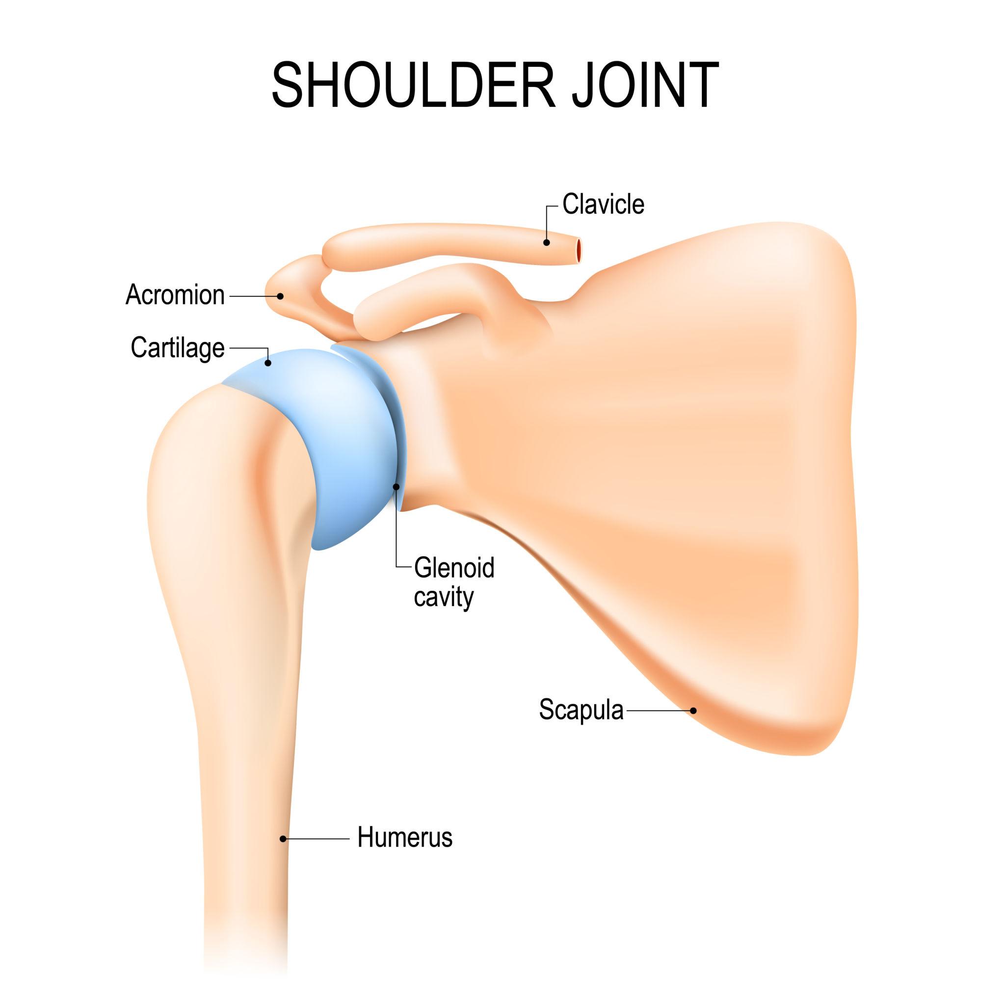
What it shows: The articulating bones: clavicle, scapula (acromion & glenoid), and humerus. The glenoid cavity is shallow, making the joint inherently unstable but highly mobile.
Importance: The glenohumeral joint is the most mobile joint in the body, but also most frequently dislocated (anterior dislocation).
Exam tip: Remember the acronym "CLAG" for structures around the shoulder: Clavicle, Ligaments (coracoclavicular), Acromion, Glenoid.
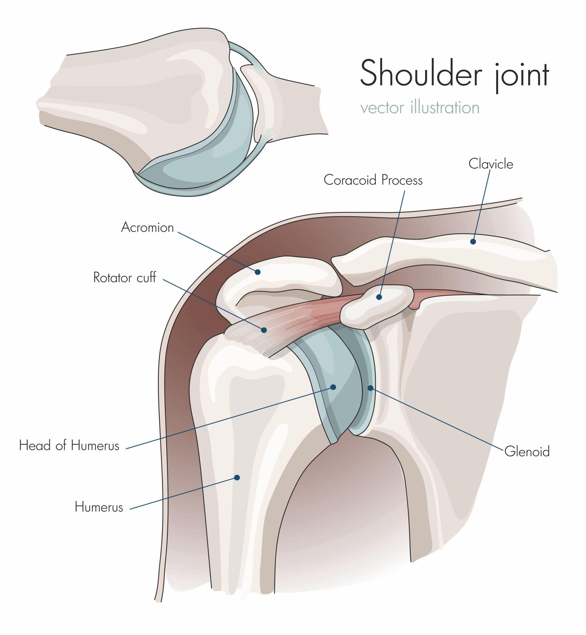
What it shows: Coracoid process, acromion, rotator cuff muscles, and head of humerus. The rotator cuff (SITS: supraspinatus, infraspinatus, teres minor, subscapularis) stabilises the joint.
Clinical correlation: Rotator cuff tears (especially supraspinatus) lead to painful arc syndrome and inability to initiate abduction.
🌀 Axilla
The axilla is a pyramidal space between the arm and thoracic wall. Contents: axillary artery (3 parts), axillary vein, brachial plexus cords, lymph nodes, and fat.
Boundaries: Apex (cervico‑axillary canal), base (skin), anterior wall (pectoralis major/minor), posterior wall (subscapularis, teres major), medial wall (serratus anterior), lateral wall (intertubercular groove).
🧠 Brachial Plexus
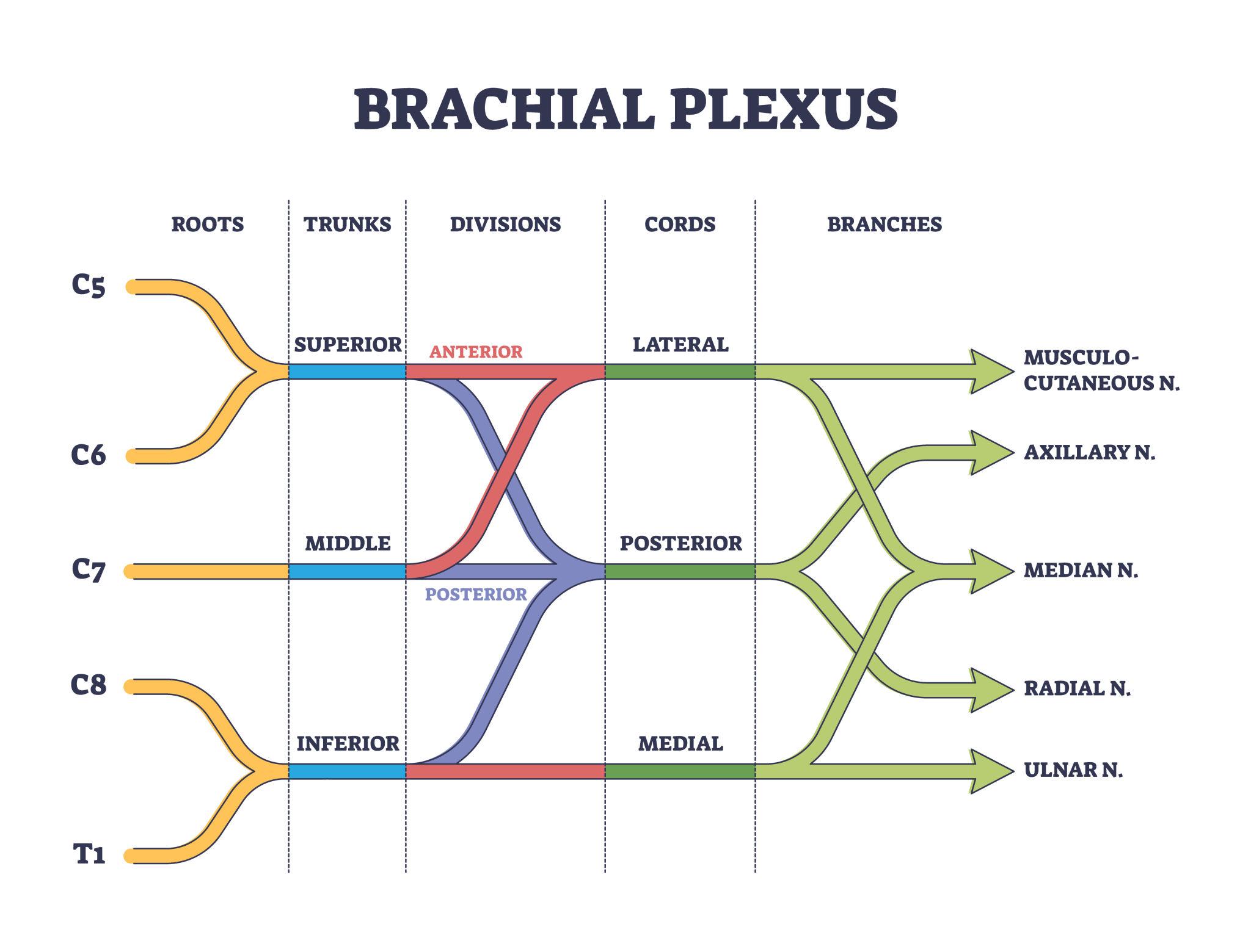
What it shows: Roots C5‑T1 → three trunks → anterior/posterior divisions → three cords → terminal nerves (musculocutaneous, median, ulnar, radial, axillary).
Mnemonics: "Randy Travis Drinks Cold Beer" – Roots, Trunks, Divisions, Cords, Branches.
Clinical pearl: Erb’s point (C5‑C6) injury causes waiter’s tip deformity; Klumpke’s paralysis (C8‑T1) causes claw hand.
💪 Arm (Anterior & Posterior Compartments)
- Anterior (flexor) compartment: Biceps brachii, brachialis, coracobrachialis. Nerve: musculocutaneous. Action: elbow flexion, shoulder flexion.
- Posterior (extensor) compartment: Triceps brachii. Nerve: radial. Action: elbow extension.
🔻 Cubital Fossa
Triangular depression anterior to elbow. Boundaries: brachioradialis (lateral), pronator teres (medial), line between humeral epicondyles (superior). Contents (medial to lateral): Median nerve, brachial artery, biceps tendon. Superficial: median cubital vein (common venipuncture site).
🦴 Forearm
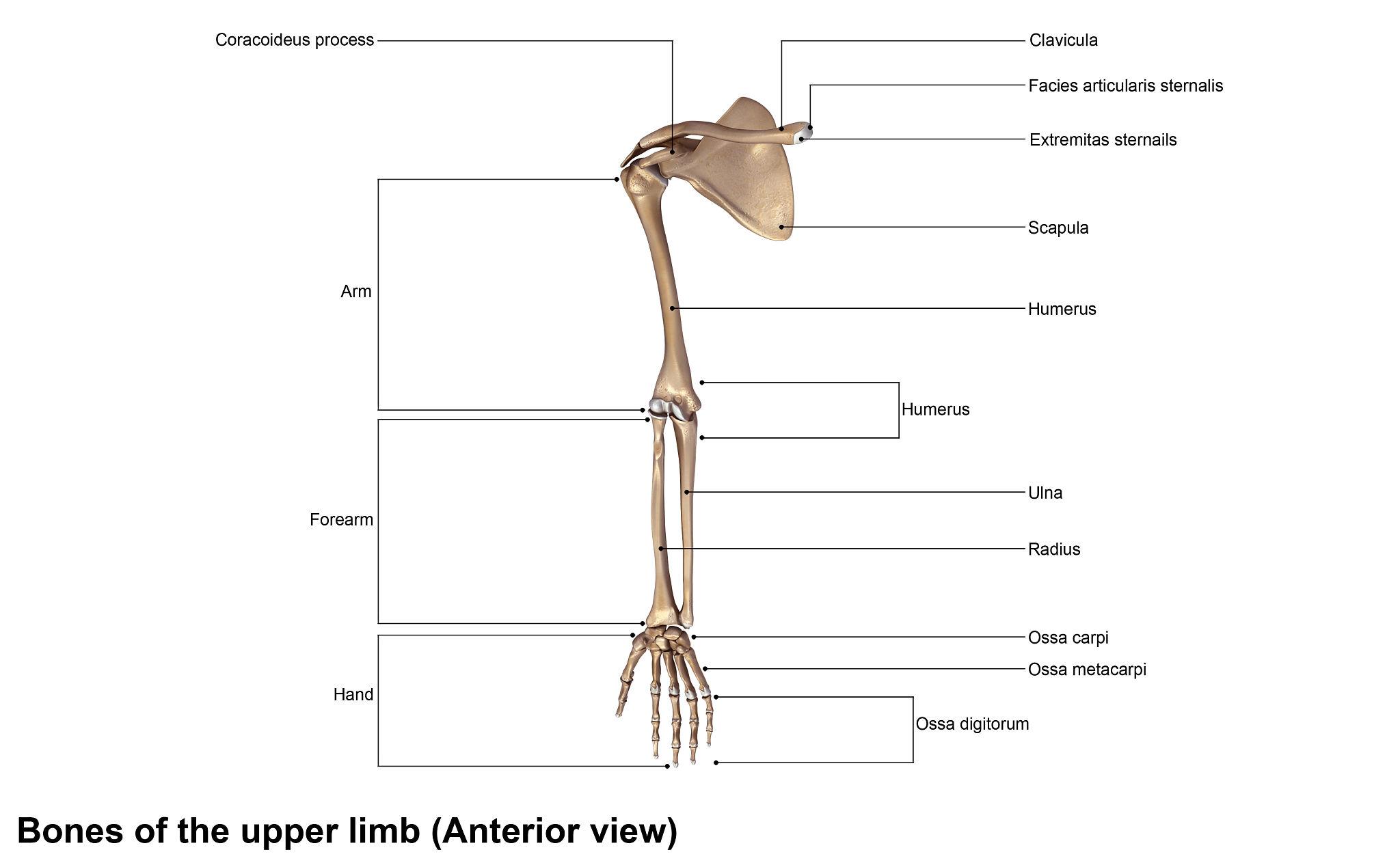
What it shows: Clavicle, scapula, humerus, radius (lateral), ulna (medial), carpal bones (8), metacarpals (5), phalanges (14).
Forearm compartments: Anterior (flexors – median/ulnar), Posterior (extensors – radial). Interosseous membrane connects radius and ulna.
✋ Wrist Joint
Condyloid (ellipsoid) synovial joint between radius and proximal carpal row (except pisiform). Movements: flexion, extension, abduction (radial deviation), adduction (ulnar deviation).
🖐️ Hand & Muscles of Hand
Thenar muscles: abductor pollicis brevis, flexor pollicis brevis, opponens pollicis (recurrent branch of median).
Hypothenar: abductor digiti minimi, flexor digiti minimi, opponens digiti minimi (ulnar nerve).
Midpalmar: lumbricals (1‑2 median, 3‑4 ulnar), interossei (3 palmar adduct, 4 dorsal abduct) – all ulnar.
🩸 Arterial Supply
Subclavian → axillary → brachial → divides into radial and ulnar in cubital fossa. Deep palmar arch (mainly radial) and superficial palmar arch (mainly ulnar) supply hand.
🩻 Venous Drainage
Superficial: cephalic (lateral), basilic (medial), median cubital vein (in cubital fossa). Deep: venae comitantes of arteries. Basilic vein becomes axillary vein.
⚡ Nerve Injuries (Clinical)
- Radial nerve: wrist drop (loss of extensors).
- Median nerve: thenar atrophy, ape‑hand deformity, loss of opposition.
- Ulnar nerve: claw hand, loss of interossei, Froment’s sign.
- Axillary nerve: deltoid paralysis – loss of abduction (15°‑90°).
- Long thoracic nerve: winged scapula (serratus anterior palsy).
📌 High‑Yield Exam Points
- Glenohumeral joint stability depends on rotator cuff & ligaments.
- Brachial plexus: roots (C5‑T1), trunks (upper, middle, lower), divisions, cords (lateral, posterior, medial), branches.
- Axillary artery has 3 parts relative to pectoralis minor.
- Cubital fossa contents: median nerve, brachial artery, biceps tendon (medial to lateral).
- Thenar muscles – median nerve; hypothenar, interossei, lumbricals 3&4 – ulnar nerve.
- Radial nerve supplies triceps and forearm extensors.
🧩 Mnemonics
Rotator cuff muscles: “SITS” – Supraspinatus, Infraspinatus, Teres minor, Subscapularis.
Carpal bones (proximal row to distal): “Some Lovers Try Positions That They Can’t Handle” – Scaphoid, Lunate, Triquetrum, Pisiform, Trapezium, Trapezoid, Capitate, Hamate.
Brachial plexus order: “Randy Travis Drinks Cold Beer” – Roots, Trunks, Divisions, Cords, Branches.
Muscles of thenar eminence: “All For One” – Abductor pollicis brevis, Flexor pollicis brevis, Opponens pollicis (median nerve).
❓ Frequently Asked Questions
📚 Recommended Resources
| Resource Name | Topic Covered | Link |
|---|---|---|
| Gray’s Anatomy for Students | Upper limb chapters | # |
| Netter’s Atlas of Human Anatomy | Shoulder & brachial plexus | # |
| TeachMeAnatomy – Upper Limb | Comprehensive notes | # |
| Kenhub Upper Limb videos | Muscles & nerve injuries | # |
| Acland’s Video Atlas (Upper Extremity) | Real dissection | # |
| Snell’s Clinical Anatomy | Clinical cases upper limb | # |
| First Aid for the USMLE | High‑yield upper limb | # |
🔎 Secondary Keywords: brachial plexus glenohumeral joint cubital fossa hand muscles nerve injuries upper limb axilla anatomy forearm compartments
📌 Long‑tail Keywords: rotator cuff muscles mnemonic upper limb arterial supply and venous drainage carpal bones labeled diagram radial nerve injury wrist drop
© 2026 MedEd – All images used for educational purposes.