General Anatomy: Complete MBBS Notes (Exam‑Oriented)
General Anatomy Notes for MBBS: Complete Guide with Images & Mnemonics
📖 Introduction
General Anatomy is the cornerstone of medical education. It provides the language to describe the human body and the framework for understanding all body systems. These notes integrate classic concepts with high‑yield facts, diagrams, and clinical pearls to help you excel in MBBS exams.
🗣️ Anatomical Terminology
Terms describe positions, directions, and regions:
- Superior / Inferior – above / below
- Medial / Lateral – toward midline / away
- Anterior / Posterior – front / back
- Proximal / Distal – closer to trunk / further
- Superficial / Deep – near surface / deeper
🧍 Anatomical Position
What it shows: Body erect, feet slightly apart, upper limbs by the side, palms facing forward (supinated), head and eyes forward. This is the universal reference.
Importance: All directional terms (anterior, posterior, etc.) are based on this position, regardless of actual posture. Essential for describing relationships, e.g., “the heart is posterior to the sternum”.
Exam relevance: Often asked in viva: “Define anatomical position.” Also used in radiology descriptions.
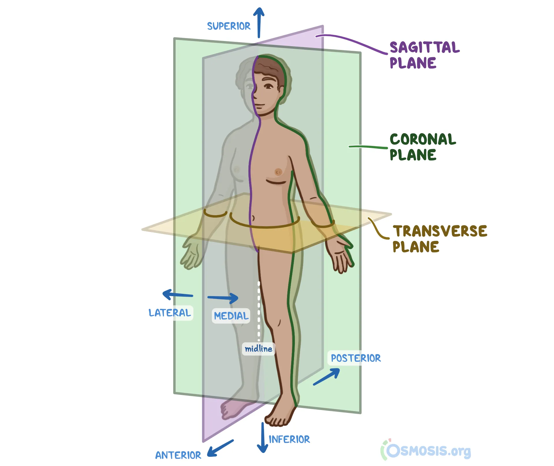
📐 Planes and Axes of the Body
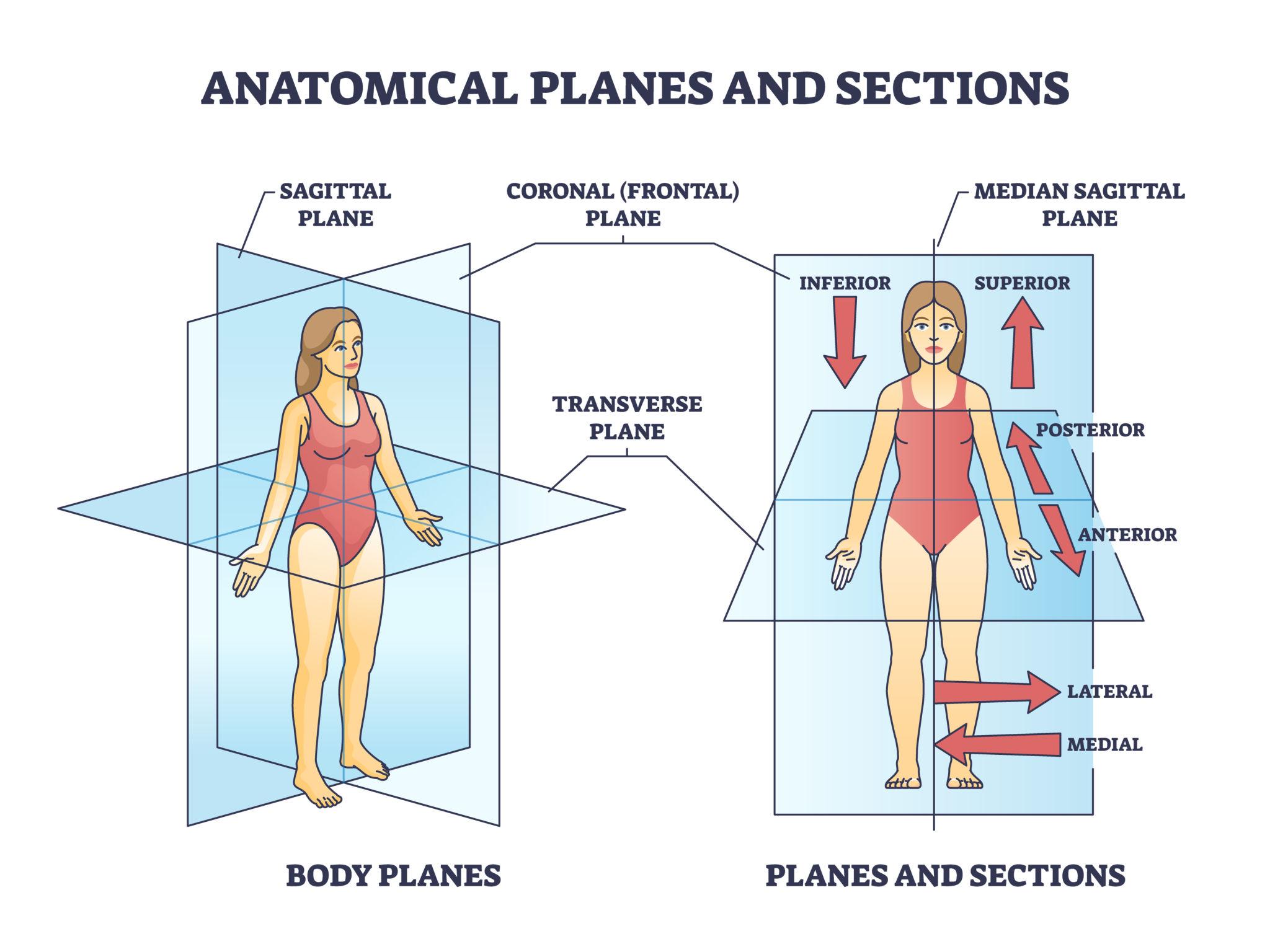
What it shows: Sagittal (median/parasagittal) divides left/right; coronal (frontal) divides front/back; transverse (axial) divides top/bottom.
Axes: Movements occur around axes perpendicular to planes. Sagittal axis → coronal plane movement; frontal axis → sagittal plane movement; vertical axis → transverse plane movement.
Clinical pearl: CT and MRI images are taken in these planes; understanding them is crucial for radiology.
🔬 Types of Tissues
Four basic tissues:
- Epithelial: covers surfaces, lines cavities (e.g., skin, gut lining).
- Connective: supports and connects (bone, blood, cartilage).
- Muscular: contracts for movement (skeletal, cardiac, smooth).
- Nervous: conducts impulses (neurons, glia).
🧪 Cell Structure Basics
Eukaryotic cell: plasma membrane (lipid bilayer), nucleus (DNA), cytoplasm with organelles: mitochondria (ATP), rough/smooth ER (protein/lipid synthesis), Golgi apparatus (modification), lysosomes (digestion).
🦴 Bones – Classification & Structure
Classification by shape: long (humerus), short (carpals), flat (sternum), irregular (vertebrae), sesamoid (patella).
Structure: diaphysis (shaft), epiphysis (ends), metaphysis (growth plate in child), periosteum (outer fibrous layer), endosteum (inner lining). Compact bone (cortex) surrounds spongy bone (trabeculae).
🧽 Cartilage
Avascular connective tissue with chondrocytes in lacunae. Types:
- Hyaline: most abundant – articular surfaces, trachea, nose.
- Elastic: ear, epiglottis – flexible.
- Fibrocartilage: intervertebral discs, menisci – resists compression.
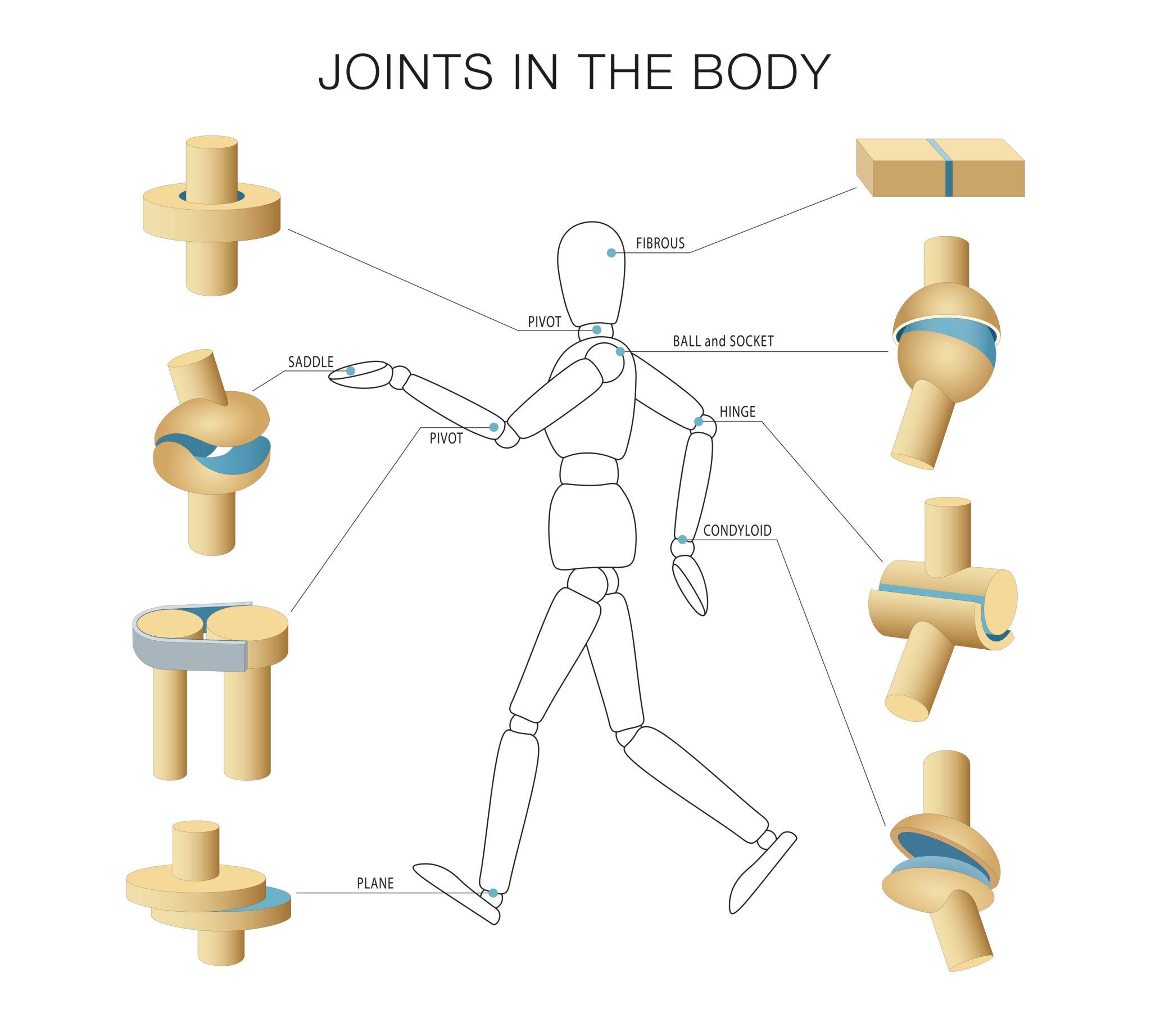
🦿 Joints – Classification & Movements
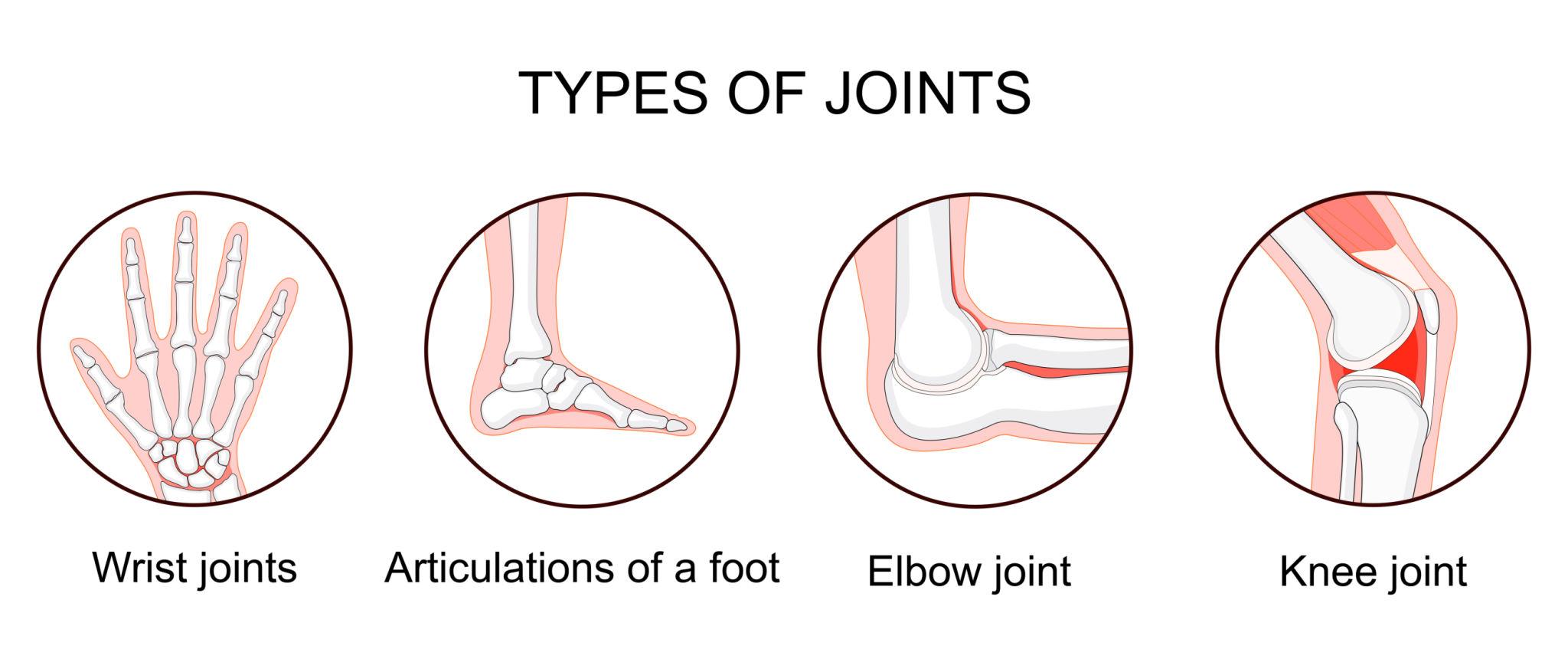
What it shows: Fibrous joints (sutures of skull) – immovable; cartilaginous (intervertebral discs) – slightly movable; synovial (knee, shoulder) – freely movable.
Functional classification: Synarthroses (fibrous), amphiarthroses (cartilaginous), diarthroses (synovial).
What it shows: Articular cartilage, joint cavity filled with synovial fluid, fibrous capsule, synovial membrane, and reinforcing ligaments.
Movements: Flexion/extension, abduction/adduction, rotation, circumduction, special moves (pronation, supination, inversion, etc.).
Exam tip: Be ready to list all six types of synovial joints: plane, hinge, pivot, condyloid, saddle, ball‑and‑socket.
💪 Muscles – Types & Physiology Basics
- Skeletal: voluntary, striated, multinucleate. Attached to bones via tendons.
- Smooth: involuntary, non‑striated, visceral organs.
- Cardiac: involuntary, striated, intercalated discs (syncytium).
Physiology basics: Sliding filament theory (actin & myosin), neuromuscular junction (ACh release), motor unit.
🩸 Blood Supply and Lymphatics
Arteries → arterioles → capillaries → venules → veins. Lymphatic system: blind‑ended capillaries, lymph nodes, trunks, and ducts (right lymphatic duct & thoracic duct). Lymph drains interstitial fluid and transports fat (lacteals).
🧠 Nervous Tissue Basics
Neurons: cell body, dendrites (receive), axon (transmit). Glial cells: astrocytes (BBB), oligodendrocytes (myelin CNS), Schwann cells (PNS), microglia (immune). Synapse – electrical or chemical.
⚙️ Mechanism: How structures work together
At a synovial joint, muscles contract, pulling tendons, causing lever‑like movement. Articular cartilage reduces friction, synovial fluid nourishes. Ligaments guide motion and prevent dislocation. Nerves (proprioception) coordinate movement.
Example: Flexion at elbow – biceps brachii and brachialis contract; triceps relax. The ulna pivots around humerus (hinge joint).
🏥 Clinical Correlation
- Dislocation: Shoulder (glenohumeral) most common anterior dislocation.
- Osteoarthritis: Degeneration of articular cartilage → bone rubbing.
- Bursitis: Inflammation of bursa (e.g., prepatellar bursitis).
- Fracture healing: Hematoma → callus (cartilage & bone) → remodeling.
- Lymphoedema: Blocked lymphatic drainage (e.g., after mastectomy).
📌 High‑Yield Exam Points
- Anatomical position → palms forward (supination).
- Three planes: sagittal, coronal, transverse.
- Four basic tissues: epithelial, connective, muscle, nervous.
- Long bone parts: diaphysis, epiphysis, metaphysis.
- Hyaline cartilage covers articular surfaces.
- Synovial joint components: cartilage, cavity, capsule, synovium, ligaments.
- Three muscle types: skeletal, cardiac, smooth.
- Lymphatic system returns interstitial fluid.
🧩 Mnemonics
Carpal bones (proximal to distal): “Some Lovers Try Positions That They Can’t Handle” – Scaphoid, Lunate, Triquetrum, Pisiform, Trapezium, Trapezoid, Capitate, Hamate.
Synovial joint types: “Planes Have Condyles, Saddles Are Ball‑and‑Socket Pivots” – Plane, Hinge, Condyloid, Saddle, Ball‑and‑socket, Pivot.
Tissue types: “Every Cell May Need” – Epithelial, Connective, Muscle, Nervous.
❓ Frequently Asked Questions
📝 Quick Revision Summary
- Position & planes: anatomical reference, 3 cardinal planes.
- Tissues & cells: 4 types, organelles.
- Bones: classification (long/short/flat/irregular/sesamoid), structure (compact/spongy).
- Cartilage: hyaline, elastic, fibrocartilage.
- Joints: fibrous, cartilaginous, synovial (6 types, components).
- Muscles: skeletal, cardiac, smooth.
- Vascular: blood vessels + lymphatics.
- Nervous: neuron + glia.
🎯 Conclusion
General Anatomy lays the foundation for every clinical subject. Mastering these basics – from anatomical position to joint classification – ensures success in exams and future bedside practice. Use the mnemonics and high‑yield points for last‑minute revision.
📚 Recommended Resources
| Resource Name | Topic Covered | Link |
|---|---|---|
| Gray’s Anatomy for Students | Comprehensive anatomy | # |
| Netter’s Atlas of Human Anatomy | Illustrations & clinical | # |
| TeachMeAnatomy | Online notes & diagrams | # |
| Kenhub | Videos & quizzes | # |
| Acland’s Video Atlas | Real cadaver dissection | # |
| Snell’s Clinical Anatomy | Clinical correlations | # |
| First Aid for the USMLE | High‑yield review | # |
| Osmosis Anatomy videos | Visual explanations | # |
🔎 Secondary Keywords: anatomical position planes of the body types of tissues bone classification joints anatomy muscle types blood supply nervous tissue basics
📌 Long‑tail Keywords: MBBS first year anatomy notes synovial joint classification and movements anatomical planes with diagrams cartilage types mnemonic
© 2026 MedEd – All images used for educational purposes.