
Complete General Embryology Notes (Week 1 to Placenta) MBBS PDF – Easy + Diagrams
Ultimate General Embryology Notes: Gametogenesis to Placenta Development
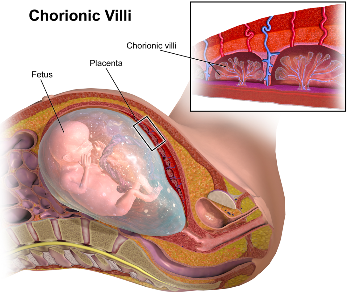
Welcome to the most comprehensive, high-yield guide on General Embryology designed specifically for MBBS 1st-year students. Instead of getting lost in heavy textbook jargon, we have simplified the entire journey of human development—from the formation of sperm and egg all the way to the development of the placenta.
Part 1: The Basics & Gametogenesis
Human development begins with the formation of gametes. These originate from Primordial Germ Cells (PGCs). PGCs are pluripotent cells produced by the epiblast during the 2nd week. They migrate to the yolk sac by the 4th week and finally reach the developing gonads by the 5th week.
Cell Potency Explained
| Type of Cell | Description | Example |
|---|---|---|
| Totipotent | Can form the entire embryo + extraembryonic tissue. | Cells up to the 8-cell stage. |
| Pluripotent | Can form all 3 germ layers (but not extraembryonic tissue). | Primordial Germ Cells, Inner Cell Mass. |
| Multipotent | Can form multiple related categories of cells. | Hematopoietic stem cells. |
Spermatogenesis vs Oogenesis
| Feature | Spermatogenesis | Oogenesis |
|---|---|---|
| Process Begins | At Puberty | During Intrauterine life (before birth) |
| Polar Body Formation | Absent | Present |
| Gametes Formed | 1 Primary Spermatocyte ➔ 4 Spermatids | 1 Primary Oocyte ➔ 1 Ovum + Polar bodies |
| Arrests (Pauses) | None | Arrests in Prophase I and Metaphase II |
Part 2: 1st Week of Development
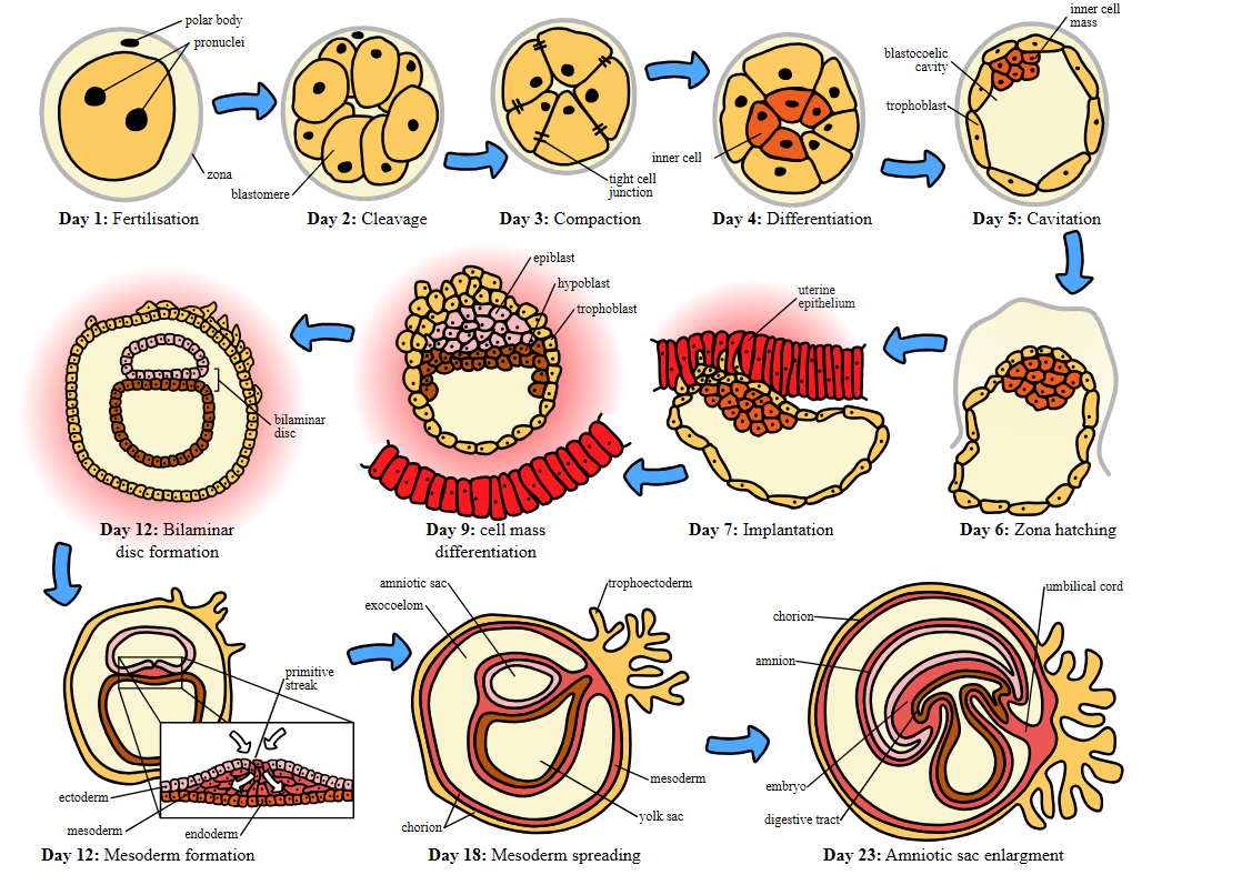
The first week is characterized by rapid cell division (cleavage) without an increase in overall size, as the conceptus travels through the fallopian tube to the uterus.
Part 3: 2nd Week - "The Rule of Twos"
During the second week, everything essentially divides into two parts.
- Trophoblast (53 cells) splits into:
- Cytotrophoblast: Inner layer.
- Syncytiotrophoblast: Outer invading layer. It produces hCG (Human Chorionic Gonadotropin), which maintains the pregnancy and is detected in pregnancy tests!
- Embryoblast (5 cells) splits into:
- Epiblast: Columnar cells (Dorsal aspect).
- Hypoblast: Cuboidal cells (Ventral aspect).
- Two Cavities form: Amniotic cavity (above epiblast) and Primary Yolk Sac (below hypoblast).
Part 4: 3rd Week - Gastrulation ("Rule of Threes")
Week 3 marks the formation of the three fundamental germ layers from the Epiblast. This process is called Gastrulation. It begins with the formation of the Primitive Streak at the tail end of the embryo.
| Germ Layer | Derived From | Major Formations |
|---|---|---|
| Ectoderm | Epiblast (Last layer to form) | Skin epidermis, Central & Peripheral Nervous System. |
| Mesoderm | Epiblast (via Primitive Streak) | Muscles, bones, blood, urogenital system. |
| Endoderm | Epiblast (Replaces hypoblast) | Lining of gastrointestinal and respiratory tracts. |
The Notochord & Mesoderm Divisions
The Notochord is a midline structure that eventually disappears, leaving behind the Nucleus Pulposus of intervertebral discs. A tumor from its remnants is called a Chordoma.
The mesoderm divides into three columns:
- Paraxial Mesoderm: Forms Somites (which further divide into Sclerotome for bones, Myotome for muscles, and Dermatome for dermis).
- Intermediate Mesoderm: Forms the Urogenital system (Kidneys, Gonads).
- Lateral Plate Mesoderm: Splits into splanchnopleuric (covers organs) and somatopleuric (body wall) layers.
Part 5: Development of Placenta
The placenta is formed from a Fetal Contribution (Chorionic Villi) and a Maternal Contribution (Decidua).
Stages of Chorionic Villi
- Primary Villus: Core of Cytotrophoblast covered by Syncytiotrophoblast.
- Secondary Villus: Extraembryonic mesoderm grows into the core.
- Tertiary Villus: Fetal blood vessels develop within the mesodermal core. (This is the functional unit!)
The Decidua (Modified Endometrium)
During pregnancy, the endometrium is called the decidua. It has three parts:
- Decidua Basalis: Deep to the embryo; forms the maternal part of the placenta.
- Decidua Capsularis: Covers the embryo like a capsule.
- Decidua Parietalis: Lines the rest of the uterine cavity.
📚 Explore Rankbaaz MBBS Study Materials
Frequently Asked Questions (FAQs)

 Continue with Google