Lower Limb Anatomy Notes for MBBS – Mnemonics
Lower Limb Anatomy Notes for MBBS: Complete Guide with Images & Mnemonics
📖 Introduction to Lower Limb Anatomy
The lower limb is specialized for weight‑bearing, locomotion, and maintenance of equilibrium. It consists of the pelvic girdle, thigh, leg, and foot. Understanding its bones, muscles, nerves, and vessels is essential for diagnosing and managing orthopedic and neurological conditions.
🍑 Gluteal Region
The gluteal region contains large extensor and abductor muscles of the hip. Key muscles: gluteus maximus (extension & lateral rotation), gluteus medius & minimus (abduction & medial rotation). Deep to gluteus maximus lie the short lateral rotators (piriformis, obturator internus, gemelli, quadratus femoris).
Sciatic nerve (L4‑S3) emerges below piriformis (in most cases) and supplies posterior thigh and all leg/foot muscles.
🦴 Hip Joint
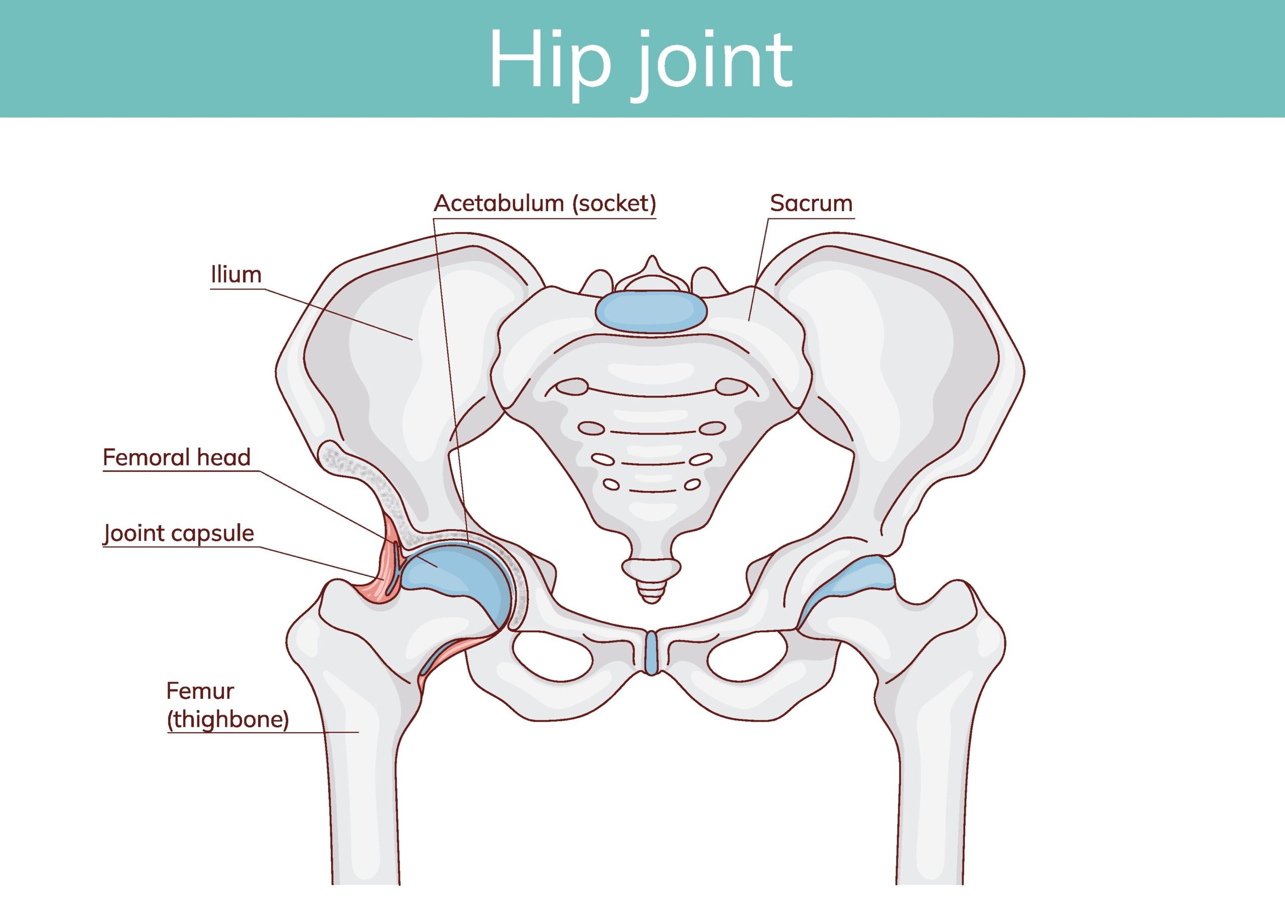
What it shows: The hip is a ball‑and‑socket synovial joint between the femoral head and acetabulum (formed by ilium, ischium, pubis). The acetabular labrum deepens the socket. The joint capsule attaches to the femoral neck.
Ligaments: Iliofemoral (Y‑ligament of Bigelow – strongest), pubofemoral, ischiofemoral. They prevent hyperextension.
Clinical note: Posterior dislocation of hip is common (from dashboard injury) and may injure sciatic nerve.
🦵 Thigh Compartments
- Anterior (extensor) compartment: Quadriceps femoris, sartorius. Nerve: femoral. Action: knee extension, hip flexion.
- Medial (adductor) compartment: Adductor longus/brevis/magnus, gracilis, obturator externus. Nerve: obturator. Action: adduction of thigh.
- Posterior (flexor) compartment: Hamstrings (biceps femoris, semitendinosus, semimembranosus). Nerve: tibial division of sciatic. Action: knee flexion, hip extension.
🔺 Femoral Triangle
Boundaries: inguinal ligament (superior), sartorius (lateral), adductor longus (medial). Floor: iliopsoas, pectineus. Roof: fascia lata, cribriform fascia. Contents (lateral to medial): Femoral nerve, femoral artery, femoral vein, empty space (lymphatics) – mnemonic: NAVEL (Nerve, Artery, Vein, Empty space, Lymphatics).
Femoral artery is a common site for arterial catheterization.
🔻 Popliteal Fossa
Diamond‑shaped fossa behind the knee. Boundaries: semimembranosus/semitendinosus (superomedial), biceps femoris (superolateral), gastrocnemius heads (inferior). Contents (from superficial to deep): tibial nerve, popliteal vein, popliteal artery (nerve‑vein‑artery). Also contains common peroneal nerve along biceps tendon.
Popliteal aneurysm may present as pulsatile mass.
🦵 Knee Joint
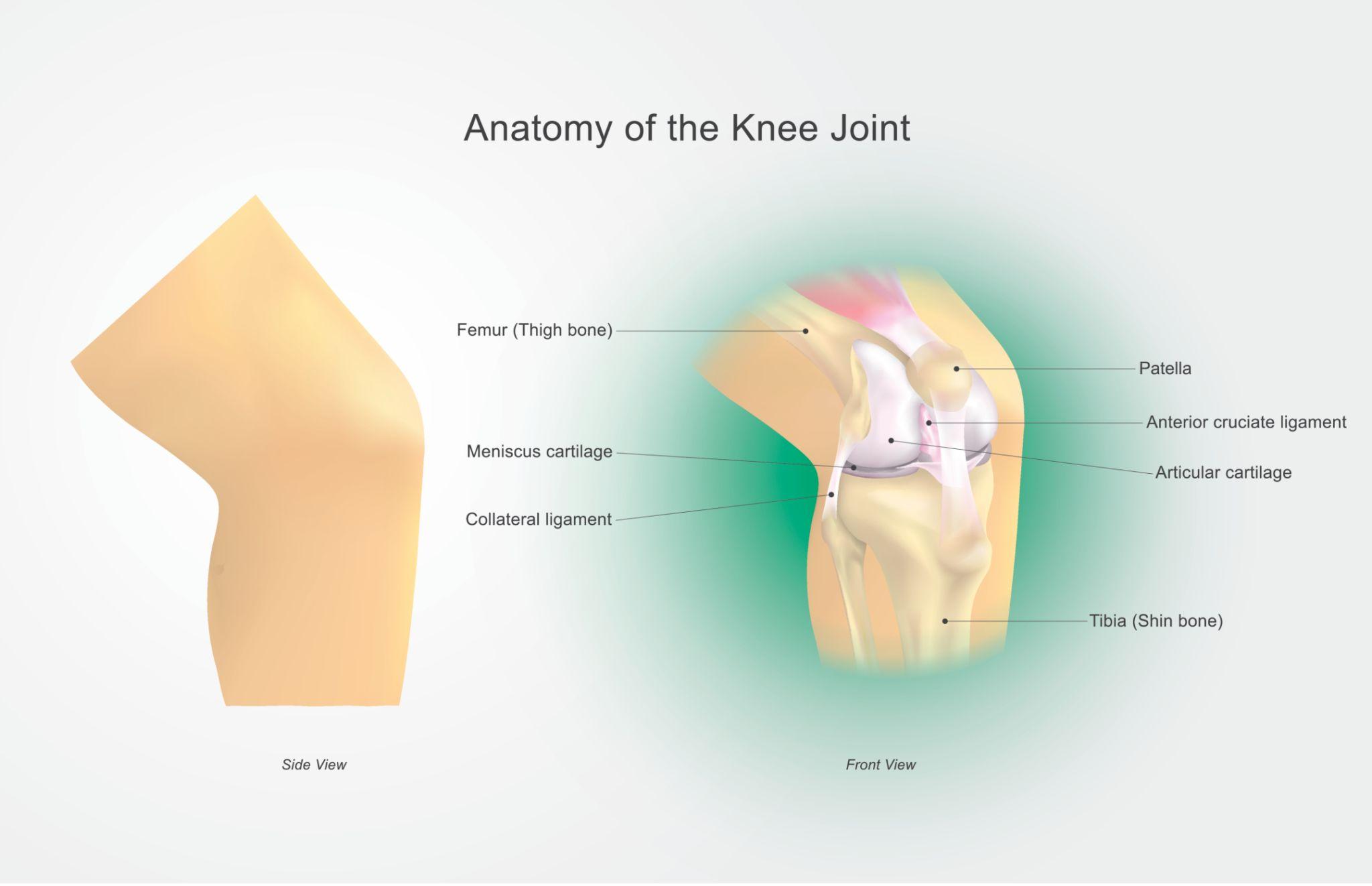
What it shows: The knee is a hinge‑type synovial joint between femur, tibia, and patella. Key features: menisci (medial and lateral) – C‑shaped fibrocartilage for shock absorption; cruciate ligaments (ACL, PCL) – prevent anterior/posterior displacement; collateral ligaments (MCL, LCL) – stabilise medially/laterally.
Clinical relevance: Unhappy triad (O’Donoghue’s triad): injury to ACL, MCL, and medial meniscus from lateral blow to knee.
Note: The second knee image URL was unavailable; we focus on this detailed diagram.
🦵 Leg Compartments
- Anterior compartment: Tibialis anterior, EHL, EDL, peroneus tertius. Nerve: deep peroneal. Action: dorsiflexion, inversion. (Structures: anterior tibial artery)
- Lateral compartment: Peroneus longus & brevis. Nerve: superficial peroneal. Action: eversion.
- Superficial posterior: Gastrocnemius, soleus, plantaris. Nerve: tibial. Action: plantarflexion.
- Deep posterior: Tibialis posterior, FDL, FHL. Nerve: tibial. Action: plantarflexion, inversion. (Posterior tibial artery)
🦶 Ankle Joint
Hinge synovial joint between tibia (medial malleolus), fibula (lateral malleolus), and talus. Movements: dorsiflexion (mainly by anterior compartment) and plantarflexion (posterior compartment). Ligaments: medial (deltoid – strong) and lateral (anterior/posterior talofibular, calcaneofibular – weak, commonly injured in inversion sprain).
🦶 Foot Anatomy
Foot bones: tarsals (7), metatarsals (5), phalanges (14). The foot is divided into hindfoot (talus, calcaneus), midfoot (navicular, cuboid, cuneiforms), and forefoot (metatarsals, phalanges).
Muscles: Intrinsic muscles – four layers of plantar muscles (abductor hallucis, flexor digitorum brevis, etc.) innervated by medial/lateral plantar nerves (branches of tibial).
🏛️ Arches of Foot
Medial longitudinal arch: Calcaneus, talus, navicular, cuneiforms, 1‑3 metatarsals. Highest point: talus. Maintained by plantar aponeurosis, spring ligament, tibialis posterior tendon.
Lateral longitudinal arch: Calcaneus, cuboid, 4‑5 metatarsals. Lower, more rigid.
Transverse arch: Runs across tarsals and metatarsal bases. Maintained by peroneus longus tendon.
Pes planus (flat foot) results from arch collapse.
⚡ Nerve Supply of Lower Limb
- Femoral nerve (L2‑L4): Anterior thigh muscles, skin of anterior thigh & medial leg (saphenous).
- Obturator nerve (L2‑L4): Adductor muscles, skin of medial thigh.
- Sciatic nerve (L4‑S3): Divides into tibial and common peroneal (common fibular) nerves.
- Tibial nerve: Posterior thigh (hamstrings), posterior leg, sole of foot.
- Common peroneal nerve: Divides into deep peroneal (anterior leg, dorsum of foot) and superficial peroneal (lateral leg, most of dorsum).
- Superior & inferior gluteal nerves: Gluteal muscles.
🏥 Clinical Correlations
- Sciatica: Pain along sciatic nerve from herniated disc or piriformis syndrome.
- Foot drop: Injury to common peroneal nerve (e.g., fibular neck fracture) – loss of dorsiflexion.
- Trendelenburg sign: Weak gluteus medius (superior gluteal nerve) – pelvis drops on unsupported side.
- Femoral neck fracture: May disrupt blood supply to femoral head (medial circumflex femoral artery) → avascular necrosis.
- Ankle sprain: Most commonly inversion injury damaging anterior talofibular ligament.
- Compartment syndrome: Increased pressure in leg compartments, often anterior, requires fasciotomy.
📌 High‑Yield Exam Points
- Hip joint: ball‑and‑socket, strong ligaments (iliofemoral), blood supply from medial and lateral circumflex femoral arteries.
- Femoral triangle: NAVEL contents, floor = iliopsoas + pectineus.
- Knee: cruciate ligaments (ACL prevents anterior translation, PCL prevents posterior), menisci (medial more commonly torn).
- Popliteal fossa: boundaries, contents (tibial nerve, popliteal vein, popliteal artery).
- Leg compartments: anterior (deep peroneal nerve, dorsiflexion), lateral (superficial peroneal, eversion), posterior (tibial nerve, plantarflexion).
- Arches: medial longitudinal (spring ligament, tibialis posterior), transverse (peroneus longus).
- Nerve injuries: common peroneal → foot drop; tibial nerve → loss of plantarflexion.
🧩 Mnemonics
Femoral triangle contents (lateral to medial): “NAVEL” – Nerve, Artery, Vein, Empty space, Lymphatics.
Popliteal fossa contents (superficial to deep): “Serve And Volley Next” – Sural nerve (or small saphenous), Popliteal vein, Popliteal artery, (nothing), (next = deeper).
Tarsal bones: “Tall Centers Never Take Shots From Corners” – Talus, Calcaneus, Navicular, Medial cuneiform, Intermediate cuneiform, Lateral cuneiform, Cuboid.
Leg compartments: “Anterior, Lateral, Superficial Posterior, Deep Posterior” – order from front to back.
❓ Frequently Asked Questions
📚 Recommended Resources
| Resource Name | Topic Covered | Link |
|---|---|---|
| Gray’s Anatomy for Students | Lower limb chapters | # |
| Netter’s Atlas of Human Anatomy | Hip, knee, foot plates | # |
| TeachMeAnatomy – Lower Limb | Comprehensive notes | # |
| Kenhub Lower Limb videos | Muscles & nerve injuries | # |
| Acland’s Video Atlas (Lower Extremity) | Real dissection | # |
| Snell’s Clinical Anatomy | Clinical cases lower limb | # |
| First Aid for the USMLE | High‑yield lower limb | # |
| Orthobullets | Trauma & anatomy | # |
🔎 Secondary Keywords: hip joint anatomy knee joint ligaments femoral triangle popliteal fossa leg compartments arches of foot sciatic nerve
📌 Long‑tail Keywords: lower limb nerve supply mnemonic medial longitudinal arch maintenance common peroneal nerve injury foot drop unhappy triad knee
© 2026 MedEd – All images used for educational purposes. Some images unavailable; we recommend consulting standard atlases.